BOOK A DEMO

* Required fields
Contact Us
Technology

The latest in early detection

There is no shortage of breakthroughs in the science of artificial intelligence, but breakthroughs often lack actual staying power in medicine because encouraging early results crumble under intense scrutiny and testing with atypical scenarios. Riverain’s deep learning AI technology is FDA cleared, clinically proven, and continues to be tirelessly tested to provide real results in radiology.

The Riverain technology difference

The standard approach to building large, complex models requires large measured training sets. These high-quality medical data sets are both time consuming and expensive to collect. Many cases look similar and do not include rare cases. Riverain developed the capability to create synthetic nodules automatically and place them into relevant anatomical contexts – such as next to the pleural wall or attached to a vessel. Our ClearRead suite of software was built on thousands of simulated, diverse nodules. By doing this, our software has been trained on more complete cases (including more rare cases), and tested on full training sets.

ClearRead

The ClearRead suite handles the most arduous medical interpretation tasks, including a systematic, thorough investigation of each voxel so that radiologists can focus on actual clinical decision making and improving their patients’ lives.

Artificial intelligence. Real results.

As the Chief Science Officer for Riverain Technologies, Jason Knapp leads a team of strategists and technicians merging the fundamental science of medical imaging with the ever-changing new technologies of artificial intelligence and deep learning. Listen to Jason’s insights on Riverain’s unique position within our industry.

Button to play video called - All CT Scans Are Not The Same
Button to play video called - Training AI to Find the Atypical
Button to play video called - The Power of Synthetic Simulatoon

As the Chief Science Officer for Riverain Technologies, Jason Knapp leads a team of strategists and technicians merging the fundamental science of medical imaging with the ever-changing new technologies of artificial intelligence and deep learning. Listen to Jason’s insights on Riverain’s unique position within our industry.

The difference is in the details

Reliable deep computation using machine learning

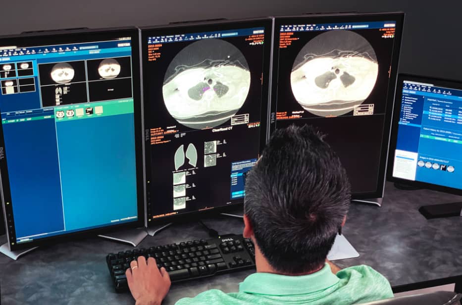
Our ClearRead technology is a modern approach utilizing the latest advances in machine learning, such as deep learning. It has surpassed the state-of-the-art by a significant margin based on a combination of frameworks, modeling, and computational ingenuity.

Achieving high reliability and significant performance usually uses substantial amounts of processing. ClearRead system can run on commodity hardware, without the need for special computer cards (GPUs) or large memory systems. By implementing this machine learning technology, our software allows radiologists to make better reads in a more timely fashion.

Acquisition independence

ClearRead handles a broad range of acquisition protocols, which is a difficult problem for automatic analysis algorithms. Riverain Technologies developed adaptive algorithms, so each study is normalized for factors such as noise, reconstruction kernels, and slice sampling effects.

Conventional approaches typically collect data from different sensors to adjust component algorithms. This leaves them vulnerable to changes in hardware, protocols, and reconstruction methods. Riverain’s adaptive process allows our software to be vendor neutral. ClearRead provides enterprise imaging without compromise, while also enabling fast and simple installation.

Reliable quantification and unique access to clinically important quantities

Vessel suppression enables improved nodule detection, but its benefit continues throughout the processing chain. Suppressing vessels and surrounding structures allows for highly precise segmentation of nodules, which provides an accurate assessment of size, volume, and other general nodule characteristics.

Vessel suppression aids the radiologist in interpretation through the removal of vascular structure within ground glass nodules, critical to the assessment of density and tissue composition. This allows a radiologist to rapidly determine the relative amount of solid tissue, which is a critical aspect for clinical decision making.